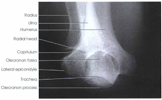
Olecranon Process - PA Axial Projection
Image Receptor: 8 X 10 inch
Patient Position
Let patient seat at the end of the radiographic table, high enough that the forearm can rest flat on the image receptor.
Part Position:
- Adjust the arm at an angle of 45 to 50 degrees from the vertical position and ensure that the patient is not leaning anteriorly or posteriorly.
- Supinate the hand and have the patient to immobilize it with the opposite hand.
- Center a point midway between the epicondyles and the center of the image receptor.
- Shield gonads.
Central ray:
The central ray is perpendicular to the olecranon process to demonstrate the dorsum of the olecranon process and at a 20 degree angle to the wrist to demonstrate the curved extremity and articular margin of the olecranon process.
Structure shown:
This projection demonstrate the olecranon process and the articular margin of the olecranon and humerus.
 |
| 0 degree angulation |
 |
| 20 degrees angulation |
Evaluation Criteria:
- The following should be clearly demonstrated:
- Olecranon process in profile
- Soft tissue outside the olecranon process
- Forearm and humerus superimposed
- No rotation of the elbow.


No comments:
Post a Comment