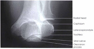
Distal Humerus - PA Axial Projection
Image Receptor:
8 X 10 inch for one or two images on 1 image receptor.
Patient Position:
Let patient seat high enough to enable the forearm to rest on the radiographic table with the arm in the vertical position. The patient must be seated so that the forearm can be adjusted parallel with the long axis of the table.
Part Position:
- Ask the patient to rest the forearm on the table, and then adjust the forearm so that the long axis is parallel with the table.
- Center a point midway between the epicondyles and the center of the image receptor.
- Flex the patient’s elbow to place the arm in a nearly vertical position so the humerus forms an angle of approximately 75 degrees from the forearm (approximately 15 degrees between the central ray and the long axis of the humerus.)
- Ensure that the patient is not leaning anteriorly or posteriorly.
- Supinate the hand to prevent rotation of the humerus and ulna, and have the patient immobilize it with the opposite hand.
- Shield gonads.
Central ray:
Central ray is perpendicular to the ulnar sulcus, entering at a point just medial to the olecranon process.
Structure shown Distal Humerus - AP Axial Projection
This radiographic projection will demonstrate the epicondyle, trochlea, ulnar sulcus (groove between the medial epicondyle and the trochlea), and olecranon fossa. This projection is used in radiohumeral bursitis or tennis elbow to detect otherwise obscured calcifications located in the ulnar sulcus.
Evaluation Criteria:
- The following should be clearly demonstrated:
- Outline of the ulnar sulcus (groove)
- Soft tissue outside the distal humerus
- Forearm and humerus superimposed
- No rotation.


No comments:
Post a Comment