Like that of adults, strategies for reducing the dose in children involve two components: one is the appropriate patient selection and appropriate technical parameters that will minimize the dose without compromising the diagnostic quality. Although radiologic technologist play an important role in dose reduction strategies for CT scanning of infants and children, it is ultimately the responsibility of the radiologist to see that such strategies are implemented.
Use CT Scan Only When Clinically Needed or Indicated
The first step in minimizing radiation exposure in children in to decide that CT scan is in fact the best method of answering the specific clinical question. Perhaps 40% of all pediatric CT scan examinations are not clearly indicated. Communication between pediatric healthcare providers and radiologist is critical in deciding whether a CT scan examination is appropriate. Radiologic Technologist play a critical role in this communication process by bringing to the radiologist’s attention any other that seems inappropriate or unnecessary. Armed with sufficient clinical information, radiologist may offer an alternative such as ultrasonography or magnetic resonance imaging (MRI) that does not use ionizing radiation. In some institutions a lack of accessibility poses a barrier to the use of other radiology imaging methods. Id modalities such as MRI are to viable alternatives that must be as easy to schedule as CT examinations. If it is decided that CT is needed the best modality, the clinical information provide can help in customizing the CT scan.
Customize the CT examination
One way of tailoring the examination to the specific diagnostic need is for the radiologist to limit the examination to the region in question. Like for example, the routine CT scanning of the pelvis as of an abdominal CT is not always necessary; in this way, exposure to the gonads will be reduced or eliminated. There are many potential situations wherein limited CT could be used. For example, in follow-up examinations, the region scanned could be limited to just the of interest like pseudocyst, lung, and abdominal abscess.
Limiting the use of multiphase examinations is another important consideration. Essentially, every additional phase increases the radiation dose by the multiple of the total number of phases. In body CT scanning, it has been reported the multiphase scanning is used in approximately 30 percent of children, many times with 3 phases. Justification for the routine use of multiphase examinations in infants and children has been questioned. Frush reported that indications for scanning through a region more than once are few, and if the use of multiphase examination were limited, the overall radiation would be at least 15 percent lower. In the rare instance less than 3 percent of the body scan, for which multiple phase are necessary, scan parameters including length of scan, slice thickness, and the tube current should be adjusted to minimize the additional radiation received.
Technical Parameters
All of the strategies outlined earlier can also be used to reduce the dose to pediatric patients. Because of a child’s increased sensitivity to radiation, additional strategies are suggested for consideration. Again it is expected that appropriate strategies are chosen and used together to reduce the dose as much as possible without sacrificing the image quality necessary to answer the clinical questions posed. It is in the mastery of the issues associated with the adjustment of each technical factor the technologist can play a unique and vital role in limiting the radiation dose to pediatric patients.
Adjustment of mAs
Appropriate selection of mAs is important for all patients, but it is imperative in children. Patient size is the primary criteria in the selection of mAs, but the region scanned and the clinical indication that prompted the scan are also considerations.
The mAs setting selected should also be based on the region scanned. Lowe tube currents are adequate in evaluating lung parenchyma. Because bone intrinsically has high contrast, the tube current should be lowered when a bone is of primary interest.
The cost of reducing mAs below a threshold point is that the signal to noise ratio decreases. The resulting noisier images have decreased low contrast resolution. In many cases, this in image quality will affect diagnosis, but in some cases the reduction may be acceptable. Like in this example; in a child for whom a large abnormality is being evaluated, such as a retroperitoneal hematoma or an abscess, the noisier image will probably be sufficient. Therefore, the current should be adjusted for patient size, region scanned, and scan indication.
Use of Patient Shielding
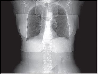
Although lead shielding is standard in general radiography, it is less beneficial in CT scan department. Because of narrow collimation, radiation to areas outside that of the selected scan area is minimal and usually attributable to the internal scattering of photons that are unaffected by surface shielding. However, a recent investigation suggested that shielding of the breast tissue and thyroid gland can be a valuable dose reduction strategy. The scout image below shows of both types of lead shielding. Perhaps equally important patient shielding may play a role in perception of risk, that is, it would assure the patient or child’s family, that every effort was being taken to reduce the radiation dose.
 |
| This scout image shows evidence of both thyroid and breast shields. These shields are a valuable dosereduction strategy. |


