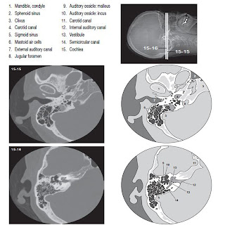
Temporal Bones – CT Study
Temporal Bone CT study this are the ct studies of the organs of hearing and balance and are located in the petrous ridge of the temporal bone. Because of these organs are tiny, thin slices are used. Once the scan data are acquired, the 2 petrosal bones are reconstructed separately so that the display field of view can reduced to ensure optimal resolution. Most departmental protocols include scans in both the coronal and axial planes. The use of intravenous contrast material varies according to the clinical indication.


No comments:
Post a Comment