MRI Arthrography
Magnetic resonance arthrography is an important
technique, especially in the hip, shoulder and ankle. A small amount of
positive contrast agent a gadolinium maybe, is injected directly to the joints.
MR arthrography is an intra-articular use of gadolinium
to diagnose rotator cuff tears, glenoid labral disruption and chondral defects.
The procedure is usually involves injecting a very dilute solution of contrast in
saline at a ratio of 1:100 or a very thin concentration of gadolinium into the
joint capsule under fluoroscopic control followed by conventional magnetic
resonance imaging. Alternatively, saline injection followed by fat suppressed
T2 weighted FSE sequence, or examining the joint after prolonged exercise to
exacerbate a joint effusion may be effective in shoulder MR arthrography.
Direct MR Arthrography
Direct MR arthrography is sometimes used in the ankle to
identify ligament tears and for increasing the sensitivity for ankle
impingement syndromes. It also has a role in assessing the stability of
osteochondral lesions and delineating loose bodies.
Indirect MR Arthrography
arthrography involves the administration of intravenous
diluted gadolinium and is sometimes used when direct arthrography is not
feasible. Although indirect MR arthrography has some disadvantages
when compared with direct MR arthrography, it does not require
fluoroscopic guidance or invasive joint injection. It is also superior
to non contrast MR imaging in delineating structures when there is minimal
joint fluid. In addition, vascularized or inflamed tissue enhances with
this method.
Temporomandibular Joints (TMJ) MRI
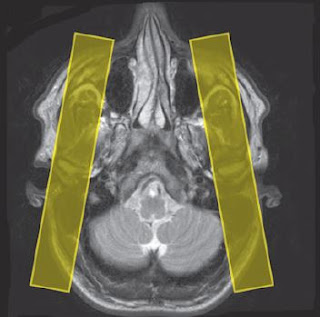
Contrast is not commonly used in this area. However,
arthrography of the joints may prove useful in the future. The joint is
injected with a small amount of gadolinium followed by sagittal/oblique T1
weighted imaging.
Elbow MR Arthrography
In elbow Arthrogram Contrast may be useful for
visualizing some soft tissue abnormalities.
In addition, elbow MR arthrography is useful for visualizing
partial- and fullthickness tears of the collateral ligament and delineating
bands in the elbow.
 |
| Coronal Elbow MR Arthrogram |
Wrist MRI
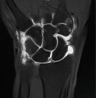
Contrast is not routinely used in the wrist; however MR
arthrography is sometimes useful in increasing the certainty of seeing
perforations of the ligaments and triangular fibrocartilage.
 |
| Wrist MR arthrogram |
Hips MR Arthrogram
IV contrast is virtually never indicated in hip joint
imaging unless as an indirect MR arthrography technique (see Shoulder in Upper
Limb). Direct MR arthrography of the hip is an important technique in the
diagnosis of some hip disorders, especially labral tears.
 |
| Hips Axial MR Arthrogram. |
Knee MRI Arthrogram
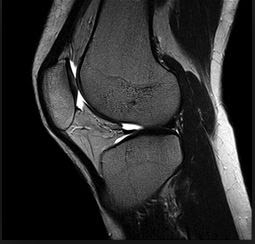
IV contrast is virtually never indicated in knee joint
imaging although it may assist in the classification of some pathologies. Knee MR
arthrography is used for the diagnosis of meniscal tears and chondral defects
and for identifying residual or recurrent tears in the knee following
meniscectomy. It also has a role in identifying loose bodies within the joint.
A very dilute solution of gadolinium in saline (1 : 100) is introduced into the
joint capsule and the single joint is imaged at high resolution with
fatsuppressed
T1 weighted images in three planes aligned relative to
the joint as described.
 |
| Knee Arthrography |
Ankle MRI Arthrography
IV contrast is not used to assess joint disease although
it may be useful for the classification of tumours. Direct MR arthrography is
sometimes used in the ankle to identify ligament tears and for increasing the
sensitivity for ankle impingement syndromes. It also has a role in assessing
the stability of osteochondral lesions and delineating loose bodies.
 | |
| Ankle Arthrography |


No comments:
Post a Comment