Hip MRI Procedure and Planning
When patient under go MRI of the hips with a prosthesis he or she may experience warmth during the scan. Inform them that this may occur and provide them with a emergency bell in case any discomfort is noticed.
MRI of the hip is usually done to evaluate these common
indications
- Evaluation of unexplained unilateral or bilateral hip pain.
- Suspected occult fracture.
- Muscle tears.
- Labral tears, chondral damage or other joint soft tissue pathology.
Bilateral and unilateral examinations of the hips are
described in this section. The causes of generalized hip pain include AVN,
metastatic deposits and occult fractures, which may affect both hips. Specific
unilateral joint pathologies such as suspected labral tears or chondral damagerequire
high-resolution imaging of the hip in question. However, due to the prevalence
of AVN in patients presenting with hip pain, it is advisable to include a
bilateral sequence in unilateral hip protocols.
Equipment used - Hip MRI
Bilateral Hip Imaging
- Body phased array/multi-coil array/general-purpose flexible coil/ body coil.
- Immobilization pads and straps.
- 20° wedge sponges.
- Ear plugs.
Single Hip Imaging
- Small/large flexible coil/multi-coil array/pelvis phased array/small Helmholtz pair.
- Immobilization pads and straps.
- 20° wedge sponges.
- Ear plugs.
Patient Preparation and Position
In the examination couch let the patient lie supine with
the legs straight and both feet are parallel to each other. In this position
the femoral neck’s angle is the same. Unlike in radiography, it is not
necessary to internally rotate the patient’s feet. Ensure legs are immobilized
with the use of pads and straps wrapped in both feet and this may help patient
to maintain the its position in a relax fashion.
Longitudinal alignment light is in the midline, and the
horizontal alignment light passes through the level of the femoral heads. This can
be found by palpating the femoral pulse which is usually can be found 3 cm
inferior and lateral to the midpoint of
the line joining the anterior superior iliac spine or ASIS and the pubic
symphysis. If only one hip needs to scan the FOV will be offset from isocenter
and image quality may be affected.
HIP MRI Suggested Protocol
Axial GRE T1
This is the localizer of the hip MRI if the three plane
localization is unavailable. This are thick slices of images on both side of
the horizontal light with Axial Inferior 25 mm to Superior 25 mm. Both hips should be included in the images to show the
location and alignment of the hips.
Coronal FSE T2 / STIR
The coronal T2 protocol on hips MRI are thin slices and shows an image of the lateral aspect of the greater trochanter to the articular portions of the acetabulum.
It particularly demonstrate on the image the flattening of the femoral head associated with AVN. Generally, FSE protocol is the sequence of choice but GRE sequence provide excellent visualization of cartilage.
 |
| Coronal FSE T1 image of both hips and femoral. |
Coronal SE/FSE T1
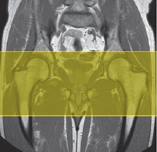
Slices are just like the same with coronal T2.Axial SE/FSE T1
Thin slices/gap are prescribed from above the articular
portion of the
acetabulum to the superior edge of the lesser trochanter,
and aligned with
the superior surface of both femoral heads to correct for
positional errors.
 |
| Coronal FSE T1 weighted image showing slice prescription boundaries and orientation for axial imaging of the hips. |
Sagittal FSE T2/coherent GRE T2* +/− chemical/spectral presaturation
Thin slices/gap are prescribed from the lateral aspect of
the greater trochanter through the articular portions of the acetabulum as shown in the image. These images particularly demonstrate flattening of the
femoral head associated with AVN. Generally, FSE is the sequence of
choice but GRE
sequences provide excellent visualization of cartilage.
 |
| Coronal FSE T1 weighted image showing slice prescription boundaries and orientation for sagittal imaging of the hips. |
Suggested protocol Unilateral MR Scanning
This examination usually demands higher resolution than
bilateral exams. Image planes should be placed relative to the anatomy of
the joint rather than orthogonal to the body. Extra shimming may be
required to optimize chemical/spectral presaturation performance and GRE image
quality in
an offset FOV.
Axial SE/FSE/incoherent (spoiled) GRE T1
Acts as a localizer if three-plane localization is
unavailable, or as a diagnostic sequence. As for bilateral examination. Use
the body coil and include both hips.
Coronal SE/FSE T1
Thin slices/gap are prescribed from the posterior to the
anterior margins of the femoral head and aligned parallel to the femoral
neck. The area from the proximal margin of the femoral shaft (below the
lesser trochanter) to the greater sciatic notch is included in the image.
Coronal coherent GRE T2*/FSE T2 +/− chemical/spectral presaturation
Slice prescription as for the Coronal T1.
T2* images are particularly good for identifying the
labrum and loose bodies in the joint. A FSE T2 may be preferred to provide
higher resolution.
Axial FSE T2 +/− chemical/spectral presaturation
Thin slices/gap are prescribed from a coronal image to
include the articular components of the hip joint. Angle the slices so that
they are parallel to the femoral neck.
Axial SE/FSE/incoherent (spoiled) GRE T1 Slice
prescription as for Axial T2.
Hip MRI Arthrogram


No comments:
Post a Comment