 |
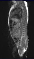
| T1 Sagital Infant MRI Lumbar |
Lumbar MRI in a Infant with Hydrocele Case
MRI of the lumbar spine with out contrast gadolinium in 1 month old infant reveals on hisMRI studies is increase in fat or fat is demonstrated inferomedial to gluteal muscles extending to the ischiorectal fossa and superolateral to the gluteus medius mucles bilaterally, but lipoma is not excluded. There is no definite leak in the CSF beyond the confines of the spinal canal. Straightening of the lumbar lordosis and a bilateral hydrocele is evidently shows on the images.
Clinical Studies Lumbar MRI
There is no definite leak of CSF beyond the confines of the visualized portions of the spinal canal.The image portion of the spinal cord appears intact. The spinal canal is not widened; there is no stenosis appreciated on the image.
The visualized portions of the lateral recesses, and neutral foramina are not compromised.
 |
| T2 sagital Lumbar MRI |
Increase in fat is also noted superior to the gluteus medius muscle, bilaterally measuring approximately 3.2 x 1.1cm, and 2.6 x 0.8cm on the right, and left side, respectively.
There is straightening of the lumbar lordosis.
The imaged vertebral bodies, their posterior elements, and the intervertebral discs apper unremarkable.
Fluid is seen in the scrotal sacs, bilaterally.
The conus medullaris terminates at the level of lumbar 1 and lumbar 2.
Download :
T1 Sagital Images of Infant Lumbar MRI
T2 Sagital Images of Infant Lumbar MRI
T2 Transverse Images Infant Lumbar MRI


No comments:
Post a Comment