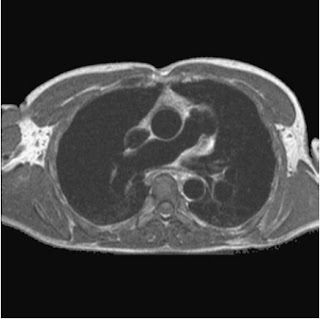
MRI the Lungs and Mediastinum
Chest MRI usually requested by doctors because to study
some common indication like:
Mediastinal lymphadenopathy
Central and superior sulcus bronchial tumors
Distinction between neoplasm and consolidated lung
Alternative to CT scan of the mediastinum and chest wall
when the patient is hypersensitive to contrast media.
Vascular evaluation of aortic dissection, pulmonary
embolus, aortic aneurysm or vascular stenosis.
Lung perfusion studies.
Assessment of diaphragmatic motion.
Equipment needed during the Chest MRI
Some common equipment like:
Body coil or volume torso multi coil array
RC bellows
ECG or peripheral gating leads
Ear plugs or headphones
Patient Positioning – MRI of the Chest
On the examination couch the patient is lying supine and
RC bellows if required, and ECG gating leads are attached. Pads are placed
under the patient’s knee and beside the patient’s elbow for optimal MR imaging.
If the patient is not comfortable supine or if the patient has trouble in
confined spaces, prone position may be an alternative position for the patient.
Patient is positioned so that the longitudinal alignment
light lies in the midline, and the horizontal alignment light passes through
the level of the 4th thoracic vertebra, or you may align it on the
patient’s nipple. To avoid unsatisfactory ECG traces the patient should place
to a feet first setting because this changes
in patient’s polarity with the main field.
Gating and Respiratory Compensation Techniques
Suggested Protocol Chest MRI
Coronal Breath Hold Fast Incoherent (Spoiled) GRE / SE T1
It acts as the localizer if the three-plane localization is not available, or it also use as a diagnostic sequence. These are medium slices and are prescribe to the vertical alignment of the lazer light, from the posterior chest muscles to the sternum. The entire lungs from apex to diaphragm or the base of the lung should be included in the image. As the chest anatomy is generally located more anterior the posterior, slice are offset in the anterior direction. |
| Localizer Chest MRI |
Axial FSE T1 or GRE T1 – Incoherent Spoiled
It is an transverse T1 view slices of the chest, except slice thickness is adjusted to fit the ROI. It is the prescribe slices from the diaphragm to the apex of the lung or through the ROI. |
|
Axial SE T1
weighted gated image of the
chest
or axial imaging.
|
Axial FSE PD / T2 / SS-FSE T2 or GRE T2
A slice prescription as for Axial T1. It is useful to characterize active tissue such as distinguishing tumors from consolidated lung and evaluate fluid pneumonia or pleural effusion. |
| Axial SE showing lesion in the left lung |


No comments:
Post a Comment