 |
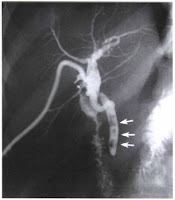
| RPO Position (AP Oblique) Postoperative Cholangiogram |
Postoperative Cholangiogram
Postoperative, Delayed, and T-tube cholangiography are radiologic terms used to the biliary tract examination that is done by way of the T-shaped tube left in the common bile duct for postoperative drainage. This examination is performed to demonstrate the caliber and patency of the ducts, the status of the sphincter of the hepatopancreatic ampulla, and the presence of remaining or previously undetected stones or other pathologic conditions.Postoperative cholangiography is performed in the radiology department. Initial or preliminary preparation usually consist of following:
 |
| Multiple Gallbladder Stones |
- The drainage tube is clamped the day before the examination to let the tube fill with bile as a preventive measure against air bubbles entering the duct, where they would simulate cholesterol stones.
- The preceding meal is withheld for at least 6-8 hrs. (NPO)
- When directed, a cleaning enema is given to patient for about 1 hour before its procedure. Premedication is not required.
- The contrast agent used is water soluble organic contrast media. Density of the contrast medium used in postoperative cholangiograms is recommended to be more than 25% to 30% because small stones may be obscured with a greater concentration.
- After an preliminary or initial radiograph of the abdomen (scot film) has been taken, the patient is adjusted in the Right Posterior Oblique (RPO) position (AP projection) with the right upper quadrant of the abdomen centered to the midline of the grid.
 |
| Location of T-TUBE (dots) |
With general precautions employed, the contrast medium is introduced und
Stern, Schein and Jacobson stressed the importance of obtaining a lateral projection is to demonstrate the anatomic branching of the hepatic ducts in this plane and to detect any abnormality that are not demonstrated. The clamp is normally not removed from the T-tube before the examination is completed. Hence the patient may be turned onto the right side for this study.
er the fluoroscopic control, spot and conventional radiographsStern, Schein and Jacobson stressed the importance of obtaining a lateral projection is to demonstrate the anatomic branching of the hepatic ducts in this plane and to detect any abnormality that are not demonstrated. The clamp is normally not removed from the T-tube before the examination is completed. Hence the patient may be turned onto the right side for this study.
are made as directed. Or else 24 x 30 cm IRs are exposed serially after each of several fractional injections of the contrast medium and then at specified intervals until most of the contrast solution has reach and entered the duodenum.


