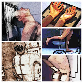
Radiographic Positioning : Radiography
The radiographic positioning terminology used by the American Registry of Radiologic Technologist (ARRT) and Canadian Association of Medical Radiation Technologist (CAMRT) are constantly the same with the following four commonterminology used in radiology:
- Projection
- Position
- View
- Method
But the only difference is that the term view is commonly used in Canada for some projections and positions.
What is Position?
What is Projection?
The term projection is distinct as the route of the central ray as it exits the x-ray tube and goes over the patient to the image receptor. Furthermost projections are defined by the entry and exit points in the body and are based on the anatomic position. AP projection for instance, are achieved when the central ray enters anywhere in the front or anterior surface of the body and exits the back or posterior. Nevertheless of which body position where patient in supine, upright, prone, and others, if the central ray pass in the anterior body surface and exits the posterior body surface the projection is called an AP projection.
Projections can also be called by the relationship made between the central ray and the body as the CR crosses through the entire body or body part like for example, the axial tangential projection.
- AP and PA Projections
- Axial Projection
- Tangential Projection
- Lateral Projection
- Oblique Projection
For additional clarification, projection may be defined by the entrance and exit points and by the central relationship to the body at the same time. In PA Axial projection for example, the central ray enters the posterior body surface and exits the anterior body surface following an axial or angled trajectory relative to the entire body or body part. Axiolateral projections also use an angulation of the central ray, but the ray enters and exits through lateral surface of the entire body or body part.
Correct patient positioning, placement of patient within the coil and proper immobilization techniques are important factors in MRI patient positioning. These are describe relatively to the light system as follow:
List of Radiographic Examinations and Positioning
- Skull
- Nasal Bone
- Chest
- Abdomen
- Upper Airway
- Upper Limbs
- Hand
- Wrist
- Scaphoid
- Forearm
- Elbow
- Humerus
- Cervical Spine
- Shoulder
- Clavicle
- Acromioclavicular Joints
- Scapula
- Toes
- Foot
- Calcaneus
- Ankle
MRI Patient Positioning
Correct patient positioning, placement of patient within the coil and proper immobilization techniques are important factors in MRI patient positioning. These are describe relatively to the light system as follow:
- The longitudinal alignment light refers to the light running parallel to the bore of the magnet in the Z axis.
- Horizontal Alignment light refers to the light that runs from left to right of the bore of the magnet in the X axis.
- The Vertical alignment light refers to the light that runs from the top to the bottom of the magnet in the Y axis
The following procedure are examined with the patient placed head first in the magnet:
 |
| MRI Laser Positioning |
- Head and Neck
- Chest
- Cervical Thoracic and Whole Spine
- Abdomen – in this examination areas superior to the iliac crest are included.
- Shoulders and UpperLimb
Anatomical Regions are examined with the patient placed Feet first in the Magnet.
- Pelvis
- Hips
- Lower Limbs
- Knee


