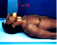
AP projection for C1 to C2 primarily demonstrating Dens
Fuchs recommends AP for demonstration of Dens if upper half is not clear in AP open-mouth. Consult the physician who reviewed the lateral cervical radiograph before attempting to move head or neck when cervical injuries is possible.Pathology demonstrated and Exposure Factors:
 |
| MML parallel to CR |
Image Receptor 18 x 24 cm or 8 x 10 inches in crosswise.
Use of grid (moving or stationary)
Patient and Part Position:
Patient is supine with the midsagital plane is center to midline of x-ray table or Image receptor.
Chin elevated to bring Mentomeatal Line (MML) near perpendicular to tabletop and adjust the angulation of central ray as needed to become parallel to MML.
Be sure no head rotation by angling the mandible equal distance to xray table.
Image receptor is centered to projected central ray.
Central ray is parallel to mentomeatal line, and inferiorly directed to the tip of mandible. Collimation and Patient Respiration:
C1 to C2 region are included to the projected collimation
Suspend patient's respiration during exposure.
Radiographic Criteria:
Structure Shown and Patient Position:
Fuchs Method AP Dens
The Dens process is centered within foramen magnum.
Symmetric appearance of mandible arched over foramen magnum evaluated as no rotation occured.
Head and neck is correctly extended evaluated if the tip of mandible clears the upper portion of the dens and the foramen magnum.
Correct Collimation and Central RayClose 4 sided collimation of C1 to C2 region.
Central Ray is to middle of dens region.
Criteria on Exposure
Proper Exposure with no motion will have clear and sharp outline of dens and other structures of C1 and C2 within the foramen magnum. For the demonstration of laminae and articular facet Cervical Spine Smith and Abel Method


