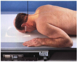
Pathology Demonstrated:
- Skull fractures (medial and lateral displacement), neoplastic processes, and Paget's diseases are demonstrated.
Technical Factors:
- IR size - 24 x 30 cm (10 x 12 inches), lenghtwise
- Moving or stationary grid
- 70 to 80 kV range
- mAs 18
Patient Position:
 |
| PA 0° CR |
- Remove all metallic or plastic objects from patient's head and neck. Exposure taken with patient in the erect or prone position.
Part Position:
- Rest patient's nose and forehead against table/bucky surface.
- Flex neck, aligning OML perpendicular to midline of table / Bucky to prevent head rotation and / or tilt (EAMs same distance from table/bucky surface).
- Center IR to CR.
Central Ray:
- CR is perpendicular to IR (parallel to OML) and is centered to exit at gabella.
- Minimum SID is 40 inches (100 cm).
Collimation:
- Collimate to outer margins of skull.
Respiration:
- Suspend respiration during exposure.
Radiographic Criteria:
Structure Shown:
 |
| PA 0° CR |
- Frontal bone, crista galli, internal auditory canals, frontal and anterior ethmoid sinuses, petrous ridges, greater and lesser wings of sphenoid, and dorsum sellae are shown.
Position:
- No rotation is evident, as indicated by equal distance bilaterally from oblique orbital to lateral margin of skull.
- Petrous ridges fill the orbits and superimpose the superior orbital region.
- Posterior and anterior clinoids are visualized just superior to ethmoid sinuses.
Collimation and CR:
- Entire skull is visualized on the image with the nasion in the center.
- Collimation borders are visible to outer borders of skull.
Exposure Criteria:
- Density and contrast are sufficient to visualized frontal bone and surrounding bony structures.
- Sharp bony margins indicate no motion.


No comments:
Post a Comment