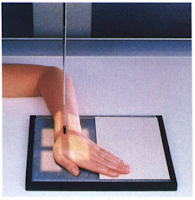
PA SCAPHOID - HAND ELEVATED AND ULNAR DEVIATION: WRIST
Warning:
- If patient has possible wrist trauma, do not attempt this position before routine wrist series has been completed and evaluated to rules out possible truama of distal forearm and/or wrist.
Pathology Demonstrate:
- Fractures of the scaphoid are shown. This is an alternative projection to the CR angle ulnar deviation method demonstrated on the preceding projection.
Technical Factors:
- IR size - 18 x 24cm (8 x 10inches)
- Division in half, crosswise
- Detail screen, tabletop
- Digital IR - use lead masking
Shielding:
- Place lead shield over patient lap to shield gonads.
Patient Position:
- Seat patient at end of table, with elbow flexed and resting on table, wrist and hands on cassette, and palm down, with shoulder, elbow, and wrist on same horizontal plane.
 |
| Ulnar Deviation |
Part Position:
- Place hand and wrist palm down on cassette with hand elevated on 20degrees angle sponge.
- Ensure that wrist is in direct contact with cassette.
- Gently evert or turn hand outward (toward ulnar side) unless contraindicated because of severe injury (fig. 5-103).
Alternative Method when Performing Scaphoid X-ray:
- Have patient clench the fist with ulnar deviation to obtain a similar position of the scaphoid.
Central Ray:
- Center CR perpendicular to IR and directed to scaphoid. (locate scaphoid at a point 2 cm [3/4 inch] distal and medial to radial styloid process.)
- Minimum SID of 40 inches (100cm)
Collimation:
- Collimate on four sides to carpal region.
Note:
- Stecher* indicate that elevation if the hands of 20degrees rather than angling of the CR places the scaphoid parallel to IR. Stecher also suggested that clenching of the fist is an alternative to elevation of the hand or angling of the CR. Bridgman: recommended ulnar deviation in addition to hand elevation for less scaphoid superimposition.
Radiographic Criteria of Scaphoid with Ulnar Deviation:
 |
| PA wrist in ulnar deviation. (5, Scaphoid; L lunate; T. triquetrum; P, pisiform; G, trapezium; M, trapezoid; C, capitate; and H, hamate.) |
Structure Shown:
- The distal radius and ulna, carpals, and proximal metacarpals are visible.
- The carpals are visible, with adjacent interspaces more open on the lateral (radial) side of the wrist.
- Scaphoid shown, without foreshortening or superimposition of adjoining carpals.
Position:
- Long axis of wrist and forearm should be align with side border of IR
- Ulnar deviation is evidence by only minimal if any superimposition of distal scaphoid.
- No rotation of wrist is evidenced by the appearance of distal radius and ulnar with no or only minimal superimposition of distal radioulnar joint.
Collimation and CR:
- Collimation should be visible on four sides to area of affected wrist.
- CR and center of collimation field should be to scaphoid.
Exposure Criteria:
- Optimal density and contrast with no motion visualize the scaphoid borders and clear, sharp bony trabecular markings.


