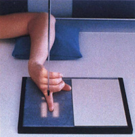
X-ray of the Fingers : Lateromedial and Mediolateral View
Lateral view of finger a fracture and /or dislocation of the distal middle, and proximal phalanges; distal metacarpal; and associated joints are shown. Some pathology, such as osteoporosis and osteoarthritis also may be demonstrated.Technical Factors
IR size - 18 x 24 cm (8 x 10 inches)Division in thirds crosswise
Detail screen, tabletop
Digital IR - use lead masking
50 to 60 kV range
Patient position, Gonadal Shielding and Accessories
Seat patient at the end of the table, with elbow flexed about 90degrees with hand and wrist resting on cassette and fingers extended.Place lead shield over patient's lap to shield gonads.
Use Sponge support block.
Lateral Projection - 2nd through 5th Digits : Part Position
 |
| Mediolateral Projection : 2nd Digit |
Align and center finger to long axis of potion of IR being exposed and to CR.
Use sponge block or other radiolucent device to support finger and prevent motion. Flex unaffected fingers.
Ensure that long axis of finger is parallel to IR.
Central Ray
CR perpendicular to IR, directed to PIP jointMinimum SID of 40 inches (100cm)
Collimation:
Collimate on four sides to affected finger.


